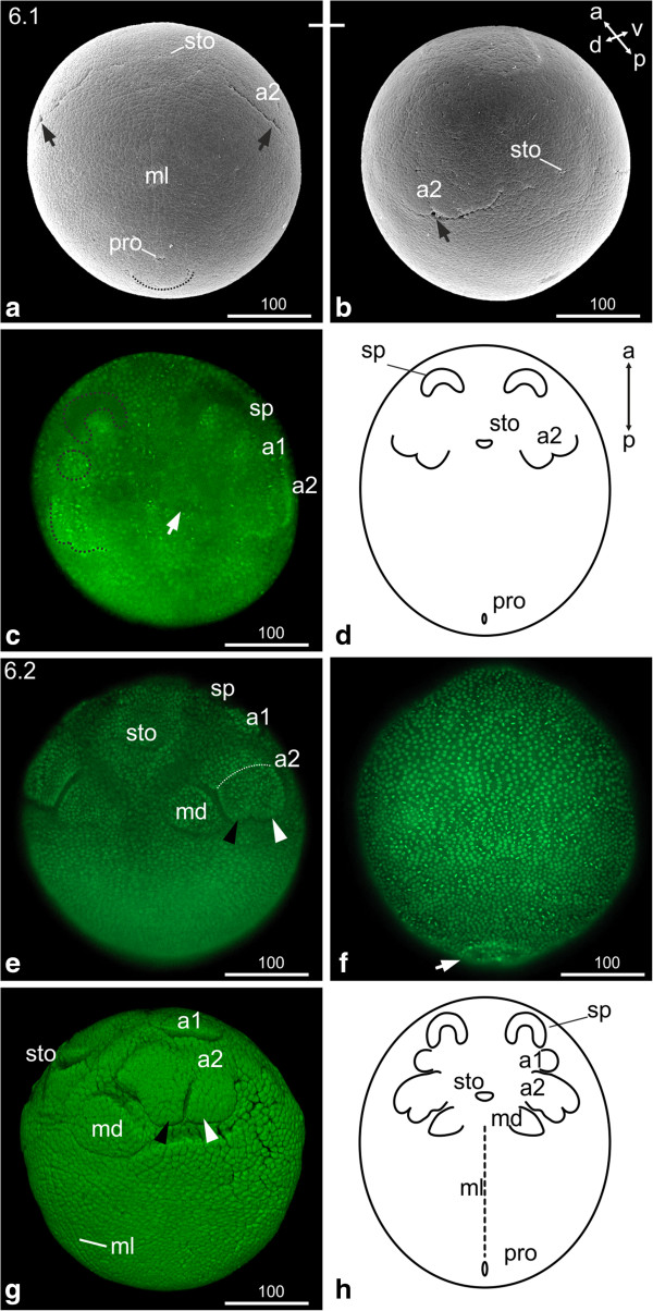
Figure 4.
Stage 6 (Naupliar segments, 6.1 (a-d); 6.2 (e-h)): (a, b) SEM; (c, e, f, g) Sytox; (d, h) Schematic drawing. (a) Stage 6.1; Ventral view: the second antennae appear as undulated transverse furrow; the arrow indicates the indentation where endo- and exopodite will form. The stomodeum appears as transverse slit. The midline cells of the developing neuroectoderm are evident. The proctodeum is visible as a ‘pore’ anterior to a ‘flap’-like structure (dashed line). (b) The same embryo as in (a) from a different view. (c) Ventral view: an area of higher nuclei density announces the appearance of the first antennae (dashed circle). The arrow points to the stomodeal area. (d) Schematic drawing of stage 6.1. (e) Stage 6.2, ventral view: the endo- and exopodal lobes of the second antennae elongate. The dashed line indicates the partition of the most proximal segment. The black arrowhead indicates the endopodite; the white arrowhead indicates the exopodite. The buds of the first antennae are evident. Mandible buds have formed medially to the second antennae. (f) Dorsal view: the ‘flap’-like incision of the proctodeal area is evident (arrow). (g) Lateral view: the buds of the first antennae are evident. The subdivision of the second antennae in to basal segment, endopodite (black arrowhead) and exopodite (white arrowhead) become clearer. The stomodeum appears as transverse slit giving the embryo a smiling appearance. The midline cells can be distinguished as longitudinal line reaching from the posterior edge of the mandibular segment to the proctodeum; they are separated via longitudinal indentations from the lateral neuroectoderm. (h) Schematic drawing of stage 6.2. a1, a2 = first and second antennae; md = mandible; ml = midline; pro = proctodeum; sp = ‘Scheitelplatte’; sto = stomodeum.

