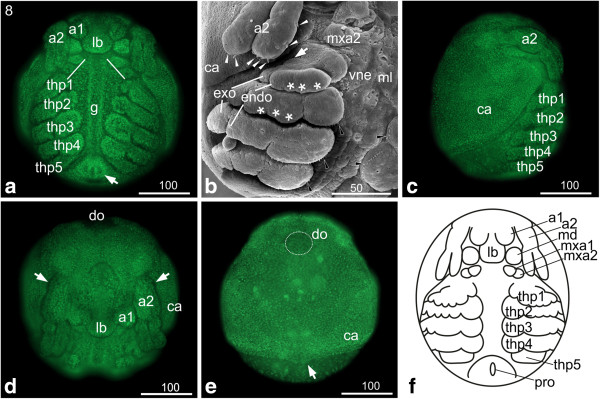
Figure 7.

Stage 8 (First antenna partially overlapping the labrum): (a, c, d, e) Sytox; (b) SEM; (f) Schematic drawing. (a) Ventral view: the flattened first antennae have continued to shift medially and partly overlap the labrum. The second antennae have elongated and reach the first thoracopod. The buds of the second maxillae have moved laterally causing the buds of maxillary appendages being lined up at a 45 degree angle relative to the anterior-posterior axis (white lines). Underneath the midline the developing gut becomes distinct. The proctodeum (arrow) has started to shift anteriorly; therefore it is positioned more medially to the fifth thoracopods. (b) Ventro-lateral view: endo- and exopodite of the second antenna show a regular arrangement of setae-buds (white arrow heads) on the tips and on the medial rim of the endopodite. The endo- and exopodites of the first to fourth thoracopod became more distinct. Tiny setae pointing to the medial food groove are visible at the margin of the basal segment of the second to fourth thoracopod (black arrowheads). (c) The lateral view shows the cape-like carapace. (d) The dorso-frontal view shows the first antenna overlapping the elongating labrum. The arrow marks the bud at the proximal segment of the first antenna. (e) Dorsal view: the carapace covers more than two thirds of the posterior area of the embryo. At the posterior edge it shows a slight medial bulge pointing posterior (arrow). The dorsal organ (highlighted by dotted line) shows a spherical shape. (f) Schematic drawing of stage 8. a1, a2 = first and second antennae; ca = carapace; g = gut; gi = gill; lb = labrum; md = mandible; mxa = maxillary appendage; pro = proctodeum; thp = thoracopod; vne = ventral neuroectoderm.
