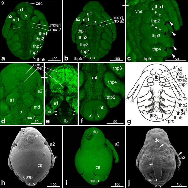
Figure 8.
Stage 9 (first and second maxillae in horizontal line): (a-f, i) Sytox; (g) Schematic drawing; (h, j) SEM. (a, b) Ventral view: the first antennae have shifted further medially, their insertions touching medially at the end of this stage. The second maxillae have moved further laterally so that they form a horizontal line with the first maxillae (dashed lines). (c) Ventro-lateral view of the thoracopods: first to fourth thoracopod continue differentiation which results in elongation of endopodites (black arrow head) and exopodites (white arrow heads) pointing postero-laterally. (d, e) In front of the compound eye anlage the nauplius eye is evident which appears in living animals as brownish eye pigment spot. The arrowheads point at the four maxillary glands in the labrum. (f) The fifth thoracopod lost its rectangular shape and shows one indentation; no setae are evident. The outgrowth of the abdomen leads to a further protruding of the proctodeum. The arrowheads indicate two developing abdominal furcal claws, which point in posterior direction. (g) Schematic drawing of stage 9. (h, i, j) The carapace protrusion narrows, becoming a short triangular process and finally a broad elongated spine. The black arrowheads in (i) and (j) point to the area where tissue movement takes place and indentations occur leading to a change in the overall-shape of the carapace. The white arrows point to the wrinkles of the embryonic cuticle. The white arrowheads in (h) and (j) point to the elongated two setae posterior of the proctodeum. (i) Shows that the dorsal organ contains only a few cells. a1, a2 = first and second antennae; ab = abdomen; ca = carapace; cec = compound eye capsule; casp = carapace spine; do = dorsal organ; lb = labrum; md = mandible; ml = midline; mxa = maxillary appendage; ne = neuroectoderm; pro = proctodeum; thp = thoracopod; vne = ventral neuroectoderm.

