Figure 9.

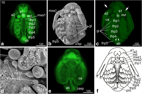
Stage 10 (Hook-shaped abdomen): (a, e) Sytox; (b, d) SEM; (c) Congo-red; (f) schematic drawing. (a) Ventral view: the second antennae have further elongated and stretch partly over the third thoracopod. (b, c) Ventro-lateral view: the abdomen appears hook-shaped due to continuous growth. The two furcal claws anterior of the proctodeum (arrowheads) have elongated and still point posteriorly. The overall shape of the thoracopods did not change, but the setae are more prominent. The fifth thoracopod just bears few tiny setae. The gills on each thoracopod have increased in size (white lines). In figure c, an egg membrane enclosing the entire embryo is evident (arrows). (d) The first maxillae have developed three prominent setae pointing medially. The second maxillae appear as small knobs showing the opening of the maxillary glands. (e) The carapace spine has developed into a narrow elongated spine, and the two halves of the carapace have assumed a seashell-like shape. (f) Schematic drawing of stage 10. a1, a2 = first and second antennae; ab = abdomen; ca = carapace; casp = carapace spine; gi = gill; lb = labrum; md = mandible; mxa = maxillary appendage; pro = proctodeum; thp = thoracopod.
