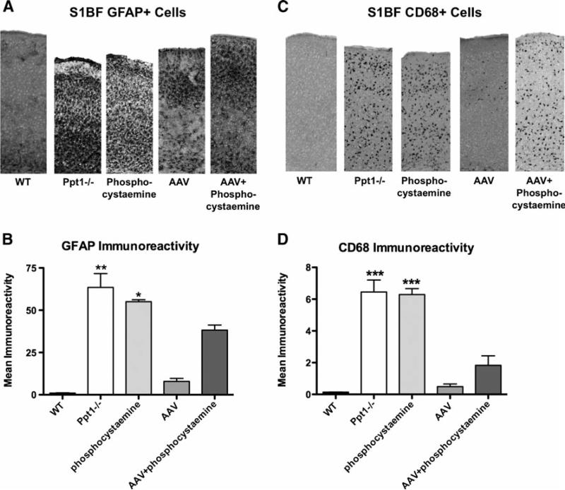
Fig. 3a-d.
Decreased neuroinflammation following therapeutic intervention. a Astrocyte activation in the S1BF cortex at 7 months. Untreated Ppt1-/-mice and phosphocysteamine-treated mice display pronounced GFAP staining in the S1BF cortex, indicative of astrocyte activation in this brain region. This was reduced following AAV only or AAV+phosphocysteamine treatment. b Quantification of GFAP immunoreactivity in the S1BF cortex at 7 months. Thresholding image analysis reveals a significant increase in GFAP immunoreacitvity in the S1BF of both untreated Ppt1-/- and phosphocysteamine-treated mice compared to WT controls. Treatment with either AAV only or AAV+ phosphocysteamine resulted in a significant decrease in GFAP immunoreactivity within the S1BF. Notably, AAV+phosphocysteamine–treated mice still displayed significant elevations in GFAP staining compared to AAV only–treated mutant mice. c Microglial activation in the S1BF cortex at 7 months. Staining for CD68 revealed a pronounced increase in staining in the S1BF cortex of Ppt1-/- mice compared to WTcontrols. A similar pattern of CD68 immunoreactivity was apparent in the phosphocysteamine-treated mice. Conversely, the S1BF of Ppt1-/- mice treated with either AAVonly or AAV+phosphocysteamine appeared to have fewer, less intensely stained CD68+ cells than were seen in untreated mutants. d Quantification of CD68 immunoreactivity in the S1BF cortex at 7 months. Both the Ppt1-/- and phosphocysteamine-treated mice displayed significantly more CD68 staining in the S1BF compared to WT controls. Relative to untreated Ppt1-/- mice, treatment with either AAV only or AAV+phosphocysteamine resulted in a significant reduction in the level of CD68 immunoreactivity in the S1BF. (*p<0.05, **p<0.01, ***p<0.001)

