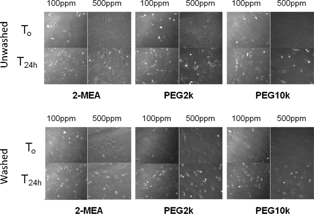
Figure 3.
Time-lapse representative images of VSMCs with 2-MEA, PEG 2K, and PEG 10K SPIONs incubation at 0 h (T0), 24 h (T24h). Cells were exposed to SPIONs either for 24 h (top: Unwashed) or 1 h and washed with media (bottom: Washed). This study was performed in DMEM at 100 ppm Fe and 500 ppm Fe of SPION concentrations.

