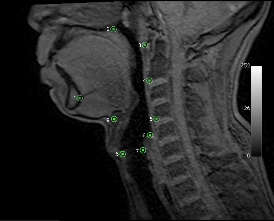
Figure 4.

Nine coordinates mapping the features of the two-sling mechanism for hyolaryngeal elevation is mapped here on a T1 weighted sagittal plane dynamic MRI. Five coordinates(1=genial tubercles of the mandible, 2=posterior edge of the hard palate, 3=anterior tubercle of C1, 4=anterior inferior edge of C2, 5=anterior inferior edge of C4) map three skeletal levers: vertebrae (line 3→4→5), mandible (line 1→3), and cranial base continuous with the hard palate(line 2→3). The next four coordinates map interconnected features of the of the hyolaryngeal complex including: 6=inferior air column of hypopharynx proximal to the upper esophageal sphincter, 7=posterior commissure of the vocal folds (posterior larynx), 8=anterior commissure of the vocal folds (anterior larynx), 9=anterior inferior edge of the hyoid.
