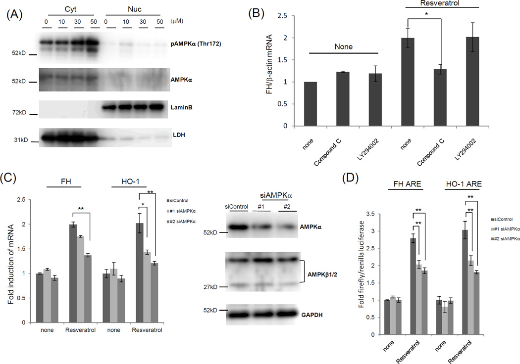
Figure 3.
Resveratrol-mediated induction of ferritin H and HO-1 mRNA is associated with AMPKα. (A) Jurkat cells were treated with different amounts of resveratrol for 2 h. Nuclear and cytosolic fractions were subjected to Western blotting with antibodies against AMPKα, phospho-AMPKα (Thr172), lamin B (nuclear marker), and LDH (cytosol marker). (B) PBMCs were pre-treated with 10 µM Compound C and 10 µM LY294002 for 1 h, followed by 30 µM resveratrol treatment for another 24 h. Total RNA was purified and subjected to quantitative RT-PCR to measure ferritin H mRNA. RT-PCR product was normalized with β-actin mRNA, and the values are indicated as a relative value of control (untreated cells). (C) Jurkat cells were transfected with 2 different siRNA against AMPKα and incubated for 48 h. Cells were treated with 30 µM resveratrol and incubated for a further 24 h. Total RNA was purified and subjected to quantitative RT-PCR to measure ferritin H and HO-1 mRNA. RT-PCR product was normalized with β-actin mRNA, and the values are indicated as a relative value of control (untreated cells). Cells were lysed and subjected to Western blotting with antibodies against AMPKα, AMPKβ1/2, and GAPDH. (D) Jurkat cells were transfected with 1 µg of human ferritin H or HO-1 ARE-luciferase plasmid along with 10 ng of pRL-null with or without siRNA against AMPKα. Cells were treated with 10 µM resveratrol for 24 h, and the resulting luciferase activity was assessed via luminometry. Fold induction was determined by setting ARE/control as 1.0. The data represent means±SEM (n≧3). *P<0.05, **P<0.01

