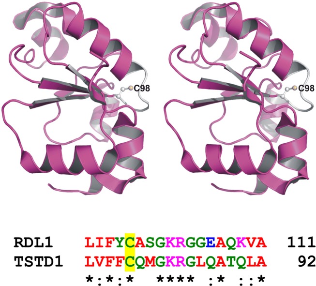
Figure 7.

Active site loop and putative catalytic cysteine in RDL1 and TSTD1. The top panel is a stereo ribbon drawing of yeast RDL1 (PDB entry 3D1P), which is shown as a magenta ribbon, except for the white active site loop. Cys98 is shown in ball and stick form. The bottom panel shows a region of a sequence alignment of RDL1 and TSTD1 around the putative catalytic cysteine (Cys98 and Cys79, respectively), which is located at the first position of the six-amino acid active site loop.
