
Figure 2.
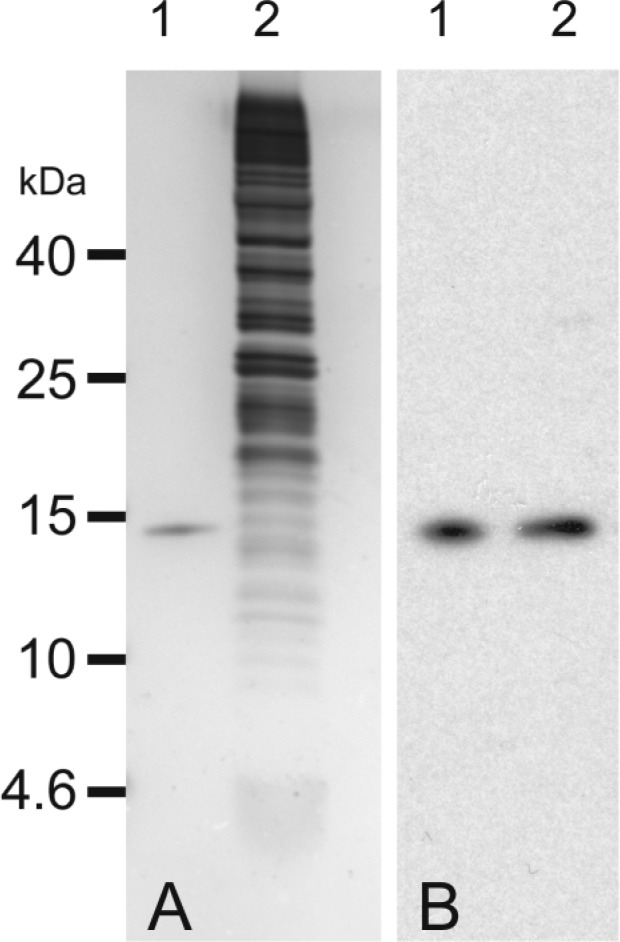
Comparison of rTHB1 and in vivo THB1: (A) 1 μg of rTHB1 (lane 1) and 1 × 10⁵ cells of CC-1690 grown in Sager-Granick M medium (lane 2) analyzed via SDS–polyacrylamide gel electrophoresis (PAGE) and stained with silver and (B) 0.5 ng of rTHB1 (lane 1) and 1 × 10⁵ cells of CC-1690 grown in Sager-Granick M medium (lane 2) separated via SDS–PAGE and transferred to nitrocellulose followed by immunodetection using purified polyclonal rabbit antibodies raised against a THB1 peptide.