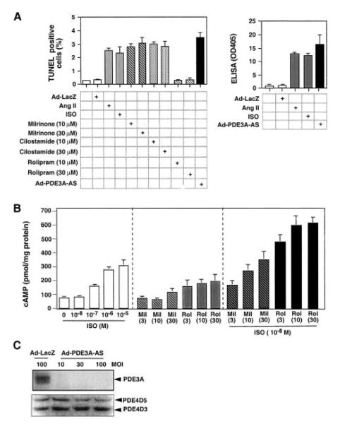
Figure 3.
Role of reduction of PDE3 or PDE4 activity in cardiomyocytes apoptosis. A, Cardiomyocytes were incubated with or without Ang II (200 nmol/L), isoproterenol (ISO) (10 μmol/L), PDE3-specific inhibitors (milrinone 10, 30 μmol/L; cilostamide 10, 30 μmol/L), or PDE4-specific inhibitor (rolipram 10, 30 μmol/L) or transduced by adenovirus vector containing antisense-PDE3A (Ad-PDE3A-AS) or LacZ (Ad-LacZ) at 30 MOI for 48 hours as indicated. Apoptosis was measured by TUNEL staining (A, left) or cell death detection ELISA kit (A, right). Absorbance at 405 nmol/L (OD 405) in sample without treatment was designated as 1.0. Data represent mean±SD of ≥4 culture preparations. B, cAMP accumulation was measured by enzyme immunoassay kit on cell lysates. Cells were treated with either various doses of isoproterenol for 5 minutes, various doses of PDE inhibitor (3 to 30 μmol/L) for 10 minutes, or PDE inhibitor for 10 minutes followed by isoproterenol (10 nmol/L) for 5 minutes. Data represent mean±SD of 3 assays. Mil indicates milrinone; Rol, rolipram. C, Western blots showing the effect of expressing PDE3A-AS via adenovirus on PDE3A and PDE4D protein expression.

