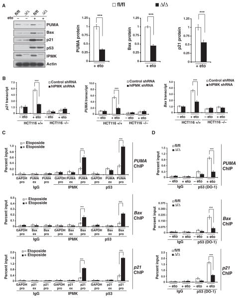
Fig. 3.
IPMK enhances p53 localization to target promoters and augments expression of p53 downstream targets. (A) Western blotting analysis of p53 and its targets PUMA, Bax, and p21 in fl/fl or Δ/Δ MEFs treated overnight with 20 μM etoposide. **P < 0.01, n = 3, mean ± SEM, Student’s t test. (B) Amounts of p21, PUMA, and Bax mRNAs in wild-type (+/+) and p53-null (−/−) HCT116 cells. ***P < 0.001, n = 3, mean ± SEM, Student’s t test. (C) Detection of IPMK at the promoters or exon regions of PUMA, Bax, and p21 by ChIP analysis of lysates from etoposide-treated wild-type MEFs. Data are means ± SEM from three experiments. ***P < 0.001, Student’s t test. (D) ChIP analysis of p53 binding to the promoter regions of PUMA, Bax, and p21 in etoposide-treated fl/fl or Δ/Δ MEFs. Data are means ± SEM from three experiments. ***P < 0.001, Student’s t test.

