Figure 2.

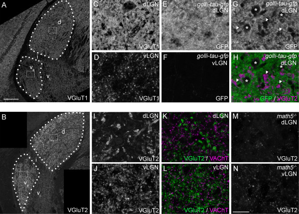
Distribution of excitatory nerve terminals in subnuclei of mouse visual thalamus. A,B: Confocal images of immunohistochemistry (IHC) for VGluT1(A) and VGluT2 (B) in coronal sections of adult mouse lateral geniculate nucleus (LGN). d, dorsal lateral geniculate nucleus (dLGN); ve, external division of ventral lateral geniculate nucleus (vLGN); vi, internal division of vLGN; i, intergeniculate nucleus (IGL). C,D: High magnification images of VGluT1-immunoreactivity in dLGN (C) and vLGN (D) from the regions boxed in A. E,F. High magnification images of Green Fluorescent Protein (GFP) distribution in dLGN (E) and vLGN (F) of adult Golli-tau-gfp transgenic mice. GFP was detected by GFP-immunostaining. Layer VI cortical neurons are selectively labeled with tau-GFP in these transgenic mice. G,H. A single optical section of a confocal image of GFP (G,H)- and VGluT2 (H)-immunoreactivity in the dLGN of an adult Golli-tau-gfp. GFP-positive cortical axon arbors densely populate the dLGN neuropil. Regions devoid of GFP-immunoreactivity in dLGN are occupied by cell bodies (asterisks), VGluT2-positive terminals (arrowheads) or blood vessels (not labeled here). I,J. High magnification images of VGluT2-immunoreactivity in dLGN (I) and vLGN (J) from the regions boxed in B. Note the difference in VGluT2-positive terminal size in dLGN and vLGN. K,L. High magnification images of VGluT2 (green) and VAChT (magenta)-containing nerve terminals in dLGN (K) and vLGN (L). VGluT2-positive terminals in dLGN are not only larger than those in vLGN, but are dramatically larger than other types of terminals in dLGN. M,N. To demonstrate that VGluT2-positive terminals originate from retinal ganglion cells, we assessed their distribution in LGN of adult math5-/- mutants, which lack retinogeniculate projections. Few, if any, nerve terminals appeared to contain VGluT2 in these mutants. All images are maximum projection confocal images except G,H. Scale bar in A = 200 μm for A,B and in N = 25 μm for C-N.
