Figure 3.

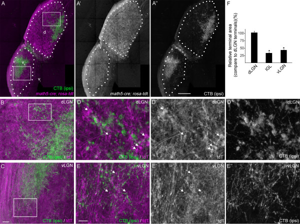
Genetic labeling of retinal terminals in subnuclei of mouse visual thalamus. A. Retinal projections (magenta) were labeled by crossing Rosa- tdt reporter mice with Math5-cre driver mice. Few (if any) cells in the thalamus express tdTomato (tdT) in Math5-cre; Rosa-tdt transgenic reporter mice. Ipsilateral retinal projections (green) were co-labeled anterogradely by intraocular injection of AlexaFluor488-conjugated cholera toxin subunit B (CTB) in the ipsilateral eye. Outlines of dorsal lateral geniculate nucleus (dLGN) and the external division of ventral lateral geniculate nucleus (vLGN) are depicted with white dots. d, dLGN; ve, external division of vLGN; vi, internal division of vLGN; i, IGL. White boxes depict regions enlarged in B and C. B,C. High magnification images of tdTomato (tdT; magenta) labeled retinal projections and CTB-labeled ipsilateral retinal projections. D, E. High magnification images of regions of dLGN (D) and vLGN (E) highlighted by the white boxes in B and C, respectively. Arrows highlight tdT-containing retinal terminals in dLGN and vLGN. All images are maximum projection confocal images. F. Relative tdT-labeled retinal terminal size (compared to retinal terminals in dLGN) was quantified in single optical sections of dLGN, vLGN and IGL. Terminal sizes in IGL and vLGN were statistical smaller than those in dLGN (P <0.001 by Neuman-Keuls Test), but were not statistically different from each other (P = 0.23). Scale bar in A = 200 μm, in C = 25 μm for B,C, in E = 10 μm for D,E.
