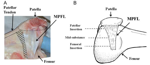
Fig. 1.

(A) A photograph showing the complex anatomy and geometry of the medial patellofemoral ligament. (B) A schematic diagram of the femur-MPFL-patella showing the fan-like shape of the MPFL with the cross-sectional area larger at the patellar attachment compared to the femoral attachment. The three designated locations where its CSA were measured are indicated.
