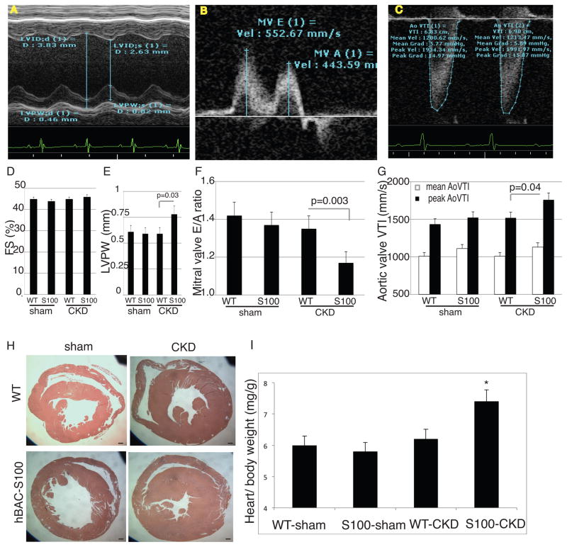
Figure 3. hBAC-S100 mice with CKD develop LVH and impaired diastolic function.
A) M-mode Doppler for measurement of fractional shortening (FS, quantified in D) and Left Ventricular Posterior Wall Thickness (LVPW, quantified in E). B) Continuous wave Doppler over the mitral valve for early (E) and atrial (A) flow was measured to calculate E/A (quantified in F). C) Continuous wave Doppler over the aortic valve was measured to calculate mean and peak aortic valve velocity timed integral (AoVTI, quantified in G). H) Representative gross pathology sections from the cardiac mid-chamber (hematoxylin and eosin stain, 5x magnification, scale bare = 20 μm) demonstrate LVH, confirmed by (I) increased ratio of heart weight to body weight in hBAC-S100/CKD mice (* P= 0.01 compared to WT/CKD and S100/sham). All values are mean ±SEM, n>20 mice per group.

