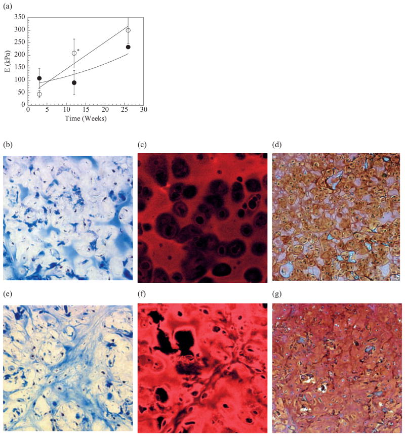
Figure 3.
Cartilagenous tissues formed via implantation of hydrogel disks containing primary bovine chondrocytes into SCID mice. The more rapidly degrading binary MVM gels led to a greater increase in the elastic modulus (E) of the engineered tissue over time, even though the MVG gels were initially more stiff a) Use of binary MVG gels for chondrocyte transplantation appeared to limit the development of new cartilage tissues over the entire time period (24 weeks) as a diffuse staining for collagen b) immunofluorescent staining for type II collagen c) and a staining for GAGs d) were observed in these tissues. In contrast, the binary MVM gels led to a higher cellularity and larger deposition of collagen e) specifically type II collagen f) and GAGs g) in the engineered tissues. * indicates differences between two conditions were statistically different (p<0.05). Tissues shown in (b) and (e) were stained with Mason’s Trichrome blue, and type II collagen in samples in (c) and (f) was visualized with a rhodamine-conjugated secondary antibody. Samples in (d) and (g) were stained with Safarin-O. All tissue sections were obtained from samples implanted for 24 weeks, and analyzed. Size bars (bar = 50 or 100 μm) are shown in figures.

