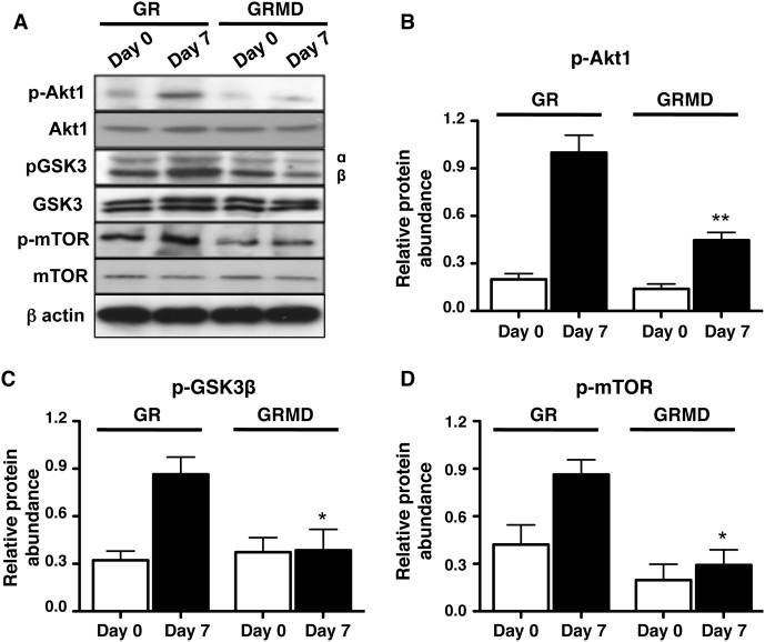
Figure 5. Loss of dystrophin reduces induction of PI3K-signaling.
For all panels, day 0 represents protein lysates obtained from serum-fed confluent cultures, and day 7 represents protein lysates obtained from confluent primary tracheal smooth muscle cell cultures (obtained from GR and GRMD animals) after 7-day serum deprivation, with medium changed every 48 h. A: representative western blots for PI3K-GSK3-mTOR signaling pathway proteins. B: densitometry analysis of the effects of serum deprivation on p-Akt1 (B), p-GSK3β (C) and p-mTOR (D) in GR and GRMD tracheal smooth muscle cells are shown. For all histograms β-actin was used as a loading control and phospho-proteins were normalized relative to their respective total protein. Data shown represent means ± SE from 6–9 experiments using 3 different primary tracheal smooth muscle cells obtained from healthy (GR) and dystrophic (GRMD) animals. Statistical comparisons shown were performed by 1-way ANOVA with Tukey’s multiple comparison tests. *p<0.05, **p<0.01, for GR day 7 versus GRMD day 7.

