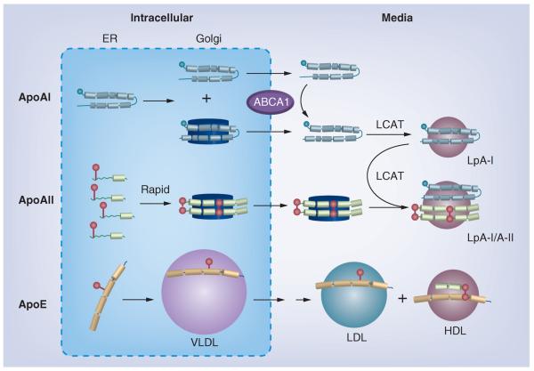
Figure 2. Hepatic HDL formation and secretion.
Apolipoprotein synthesis and its initial lipidation occur in the ER. In the Golgi, apoAII particles are further lipidated and approach the size of HDL, while apoAI particles are smaller, with more than half of the intracellular apoAI remaining lipid-free. After secretion, apoAI acquires lipid via ABCA1, and nascent apoAI HDL matures to spherical LpA-I by action of LCAT. ApoAII, which dimerizes after lipidation, is secreted as a lipidated dimer on particles without apoAI. Shortly after secretion, LCAT promotes fusion of these particles with spherical apoAI–HDL. Monomeric apoE in the Golgi associates with VLDL, which are remodeled after secretion (double arrow) to LDL-sized particles and some apoE transfers to HDL, where it dimerizes with apoAII. Apolipoproteins are colored as: gray (apoAI), green (apoAII) and orange (apoE) rods to show helix structure; cysteine groups are red spheres.
ER: Endoplasmic reticulum; Lp: Lipoprotein.
Reproduced with permission from [31].

