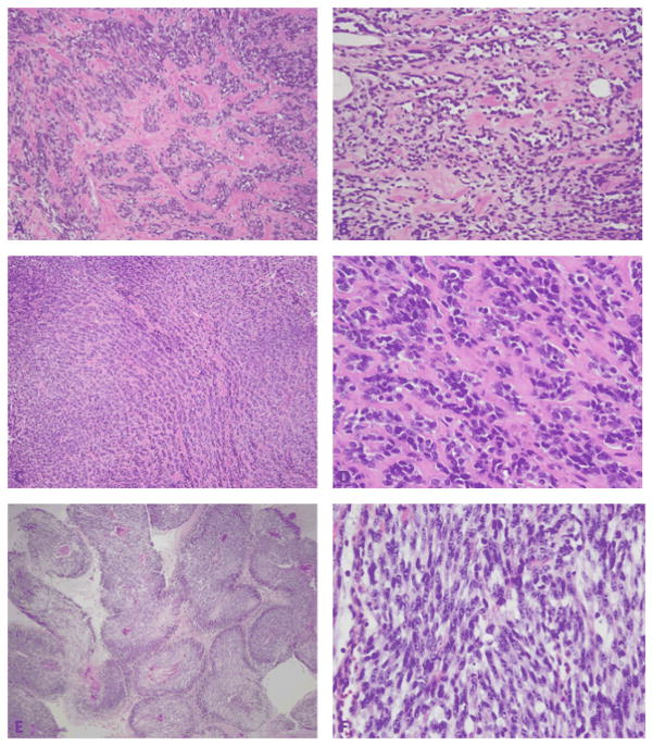
Figure 3.
Pathologic spectrum of MYOD1-mutant RMS. (A, B) Sclerosing RMS from the thigh in a 76 year-old male (Case 3, H&E 100×) showing cellular clustering in a densely sclerotic background; (B) which at higher power display a distinctive pseudo-vascular growth pattern (Case 3, H&E, 200×). (C) Sclerosing RMS in a 15 year-old girl (case 5, H&E, 100×) showing small nests of cells in a sclerotic background; (D) which at higher magnification reveal small blue round cells with minimal cytoplasm embedded in a collagenous stroma (case 5, H&E, 400×). (E) Spindle cell RMS from the buttock in a 2 year-old girl (Case 7, H&E,100×) showing perivascular distribution of viable spindle cells with intervening geographic necrosis, reminiscent of an MPNST-like morphology; (F) higher power showing elongated spindle cells with hyperchromatic nuclei arranged in a fascicular pattern (Case 7, H&E 400×).

