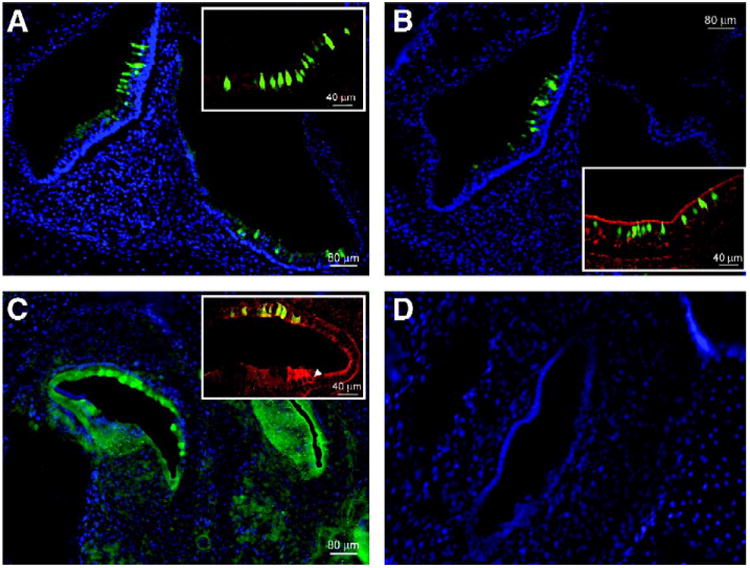
Fig. 2.

Sagittal cryosections of the murine cochlear and vestibular systems. eGFP-expressing hair cells and supporting cells are seen with fluorescence microscopy in cochlear and vestibular sagittal cryosections of P0 mice that had been injected at E12. (A) Vestibular section after exposure to AAV2/1 (original magnification 10×). The inset (original magnification 25×) shows transduced hair cells and supporting cells that express eGFP. Anti-myosin VIIa antibody is used as a hair cell marker and the reactive cells appear red. Hair cells that express eGFP as well as myosin VIIa appear green-yellow at the colocalization sites. (B) AAV2/8-injected cryosections are seen at 10× original magnification with a 25× original magnification inset. (C) Cochlear section after exposure to lentivirus and staining with an anti-eGFP antibody followed by FITC for better visualization. The white arrow in the inset indicates a myosin VIIa-stained hair cell. Nonspecific eGFP colocalization with this stain appears yellow-green. (D) Nuclei (stained blue with DAPI) outline the macula in the utricle in a sample from an AAV2/9-injected mouse. eGFP is absent.
