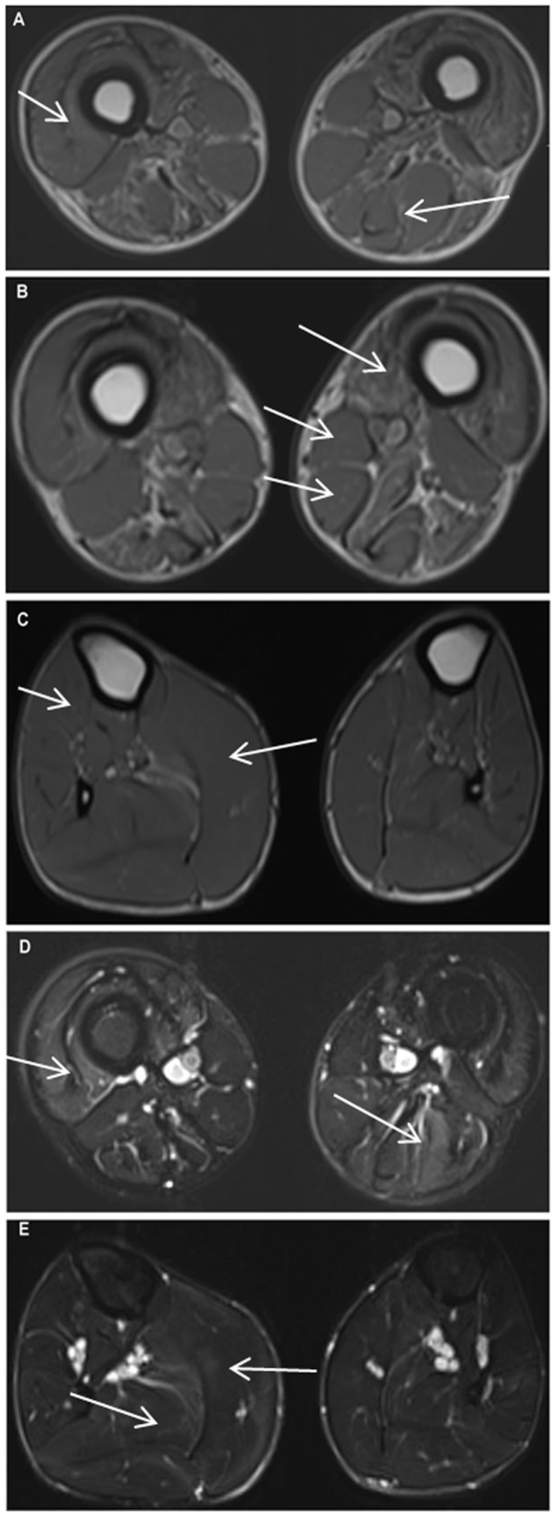
Figure 3. Muscle MRI of patient F97-1.

A. T1W image showing global atrophy of muscles of anterior compartment, medial compartment and posterior compartment of thigh. Biceps femoris long head shows stage 3 fatty infiltration. Short head of biceps femoris, sartorius and gracilis are hypertrophied; B. shows severely atrophied vastus intermedius muscle, Mercurie stage 2b infiltration of vastus medialis muscle. Short head of biceps femoris, sartorius and gracilis are hypertrophied; C. Tibilialis anterior, extensor hallucis longus and extensor digitorum longus are atrophied (right more than left). On right side the medial head of gastrocnemius is hypertrophied and lateral head is atrophied. Left soleus is atrophied. Mild fatty infiltration of soleus muscle is noted in left side. Striking asymmetry is present; D. STIR image of thigh shows asymmetrical myoedema pattern. On the right side there is stage 3a myoedema and on the left side it is stage 2a. There is stage 2b myodema noted in the posterior compartment on left side; E. Myoedema mainly seen in gastrocnemius and soleus muscle. Stage 2b on right side and stage 2a on left side.
