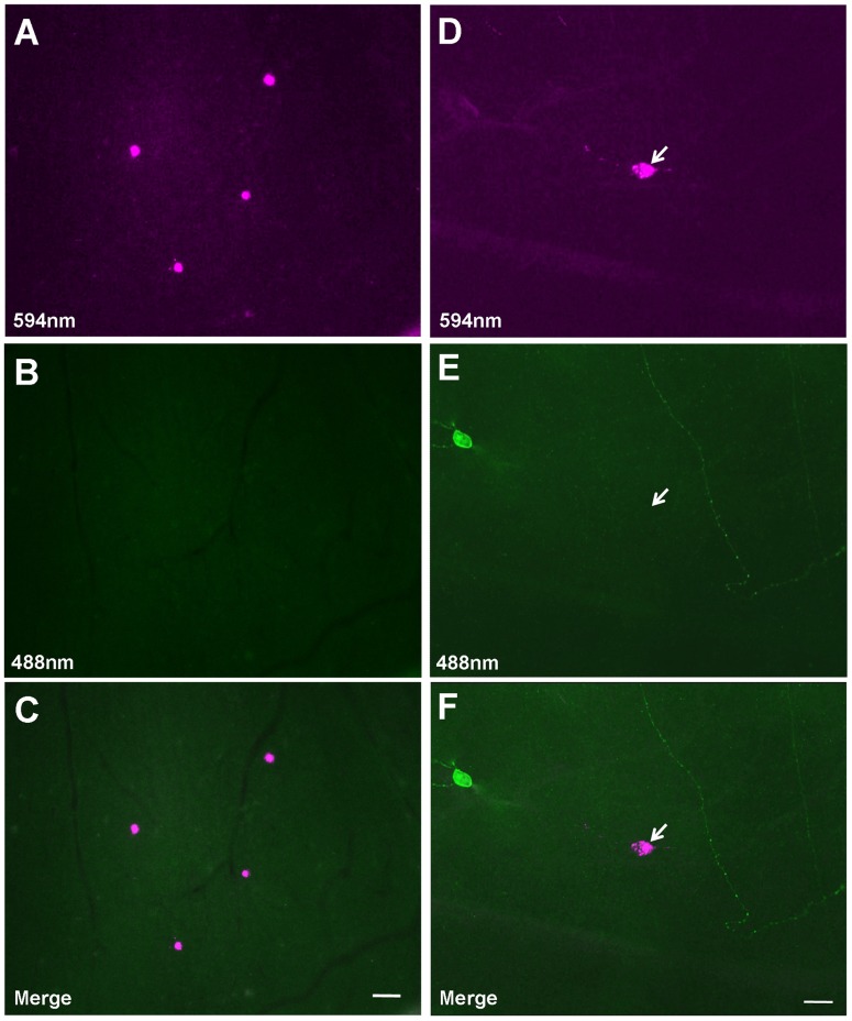
Figure 2. Illustration of Cholera toxin B subunit (CTB) retrogradely labeled-caudal periaqueductal gray (cPAG)-projecting retinal ganglion cells (RGCs).
(A–C) Illustration of the CTB-conjugated Alexa Fluor 594 (CTB-594) retrogradely labeled RGCs in the retina. There are 4 CTB-labeled RGCs (magenta) in this field (A). These cells were not visible under blue excitation florescence filter (B). Merged photomicrograph of (A) and (B) is shown in (C). (D–F) A cPAG-projecting RGC is negative for anti-melanopsin staining. Arrow points to a CTB-594 retrogradely labeled cPAG-projecting RGC (D) that, as shown in (E), lacks melanopsin immunoreactivity. In contrast, melanopsin-positive RGCs were also seen. As shown in (E), the soma and dendrites of a melanopsin-expressing RGC (mRGC) were visible, so was an dendrite of another mRGC whose soma was not in the field (green fluorescent staining). Merged photomicrograph shows a cPAG-projecting RGC and a non-cPAG-projecting but melanopsin-positive RGC in the same field (F). Scale bars: 40 µm in C (applies to A and B); 20 µm in F (applies to D and E).

