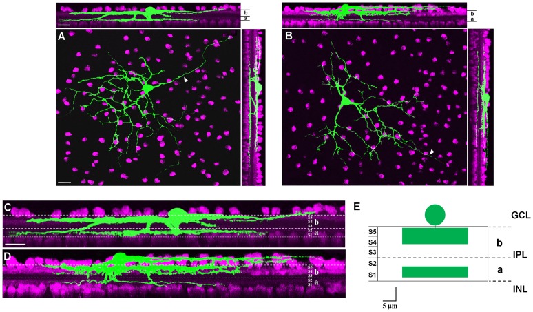
Figure 6. Dendritic stratification of caudal periaqueductal gray (cPAG)-projecting retinal ganglion cells (RGCs).
(A–B) These two cells are distinctly bistratified in sublamina a and b of the inner plexiform layer (IPL). Cholinergic amacrine cells were labeled with color magenta. (C–D) High-power images of upper planes in (A) and (B), illustrating that these two cPAG-projecting RGCs could distinctly bistratified in IPL. White dotted lines indicated borders of sublamina a and b of IPL. (E) Schematic summary of ramification pattern of cPAG-projecting RGCs. INL: inner nuclei layer; IPL: inner plexiform layer; GCL: retinal ganglion cell layer; a: sublamina a of inner plexiform layer; b: sublamina b of inner plexiform layer; S1–S5: stratum 1–5. Scale bars: 20 µm in A (applies to B); 20 µm in upper panel of A (applies to upper panel of B); 20 µm in C (applies to D); 5 µm in E.

