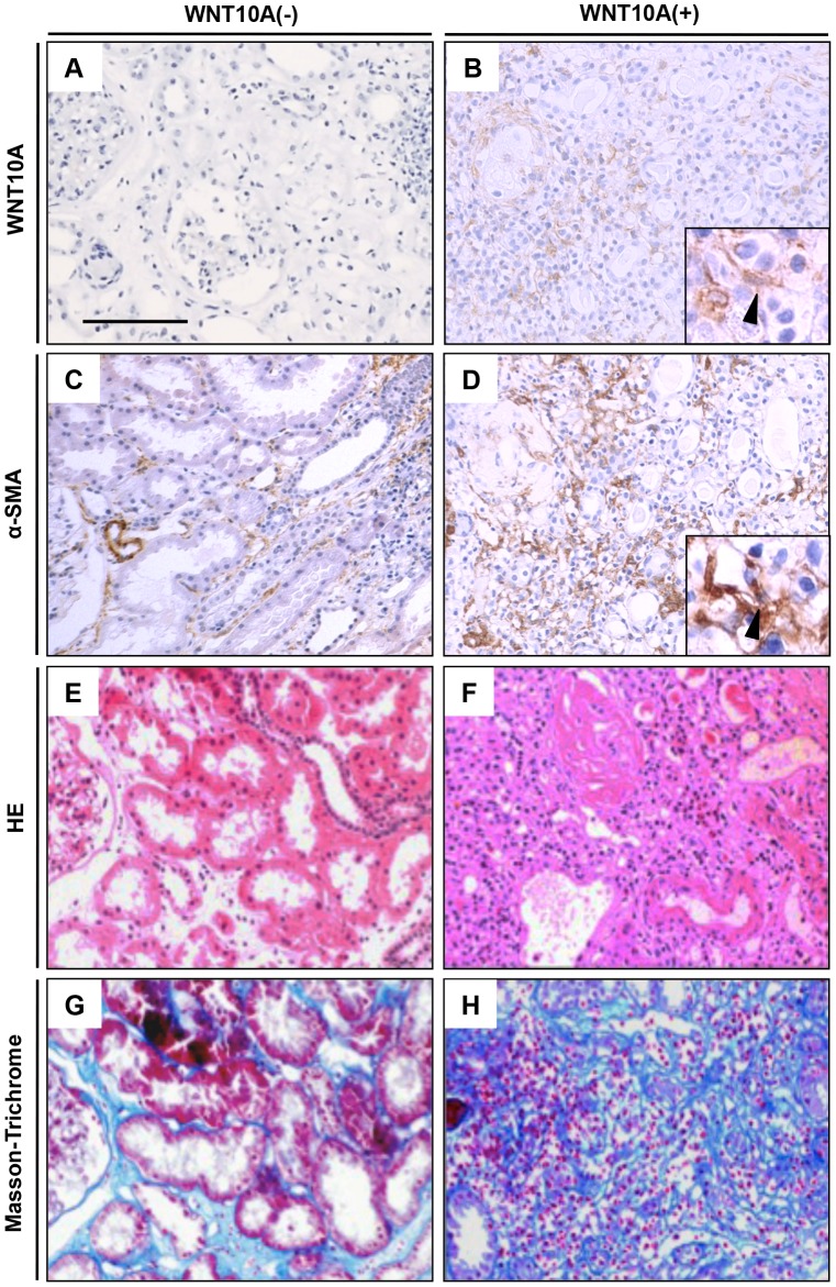
Figure 2. Histopathological analysis of kidney tissue from acute interstitial nephritis patients.
Left panels are kidney interstitium without WNT10A expression, and right panels are kidney interstitium with WNT10A expression. (A–B); immunohistochemical staining for WNT10A, (C–D); immunohistochemical staining for α-SMA, (E–F); HE staining, (G–H); Masson-Trichrome staining. Arrow head is spindle cell with WNT10A and α-SMA positive respectively. All photos were taken at 200×. Scale bar is 100 µm.

