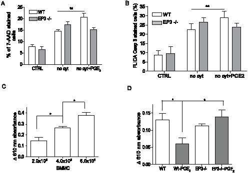
Figure 4. EP3 dependent apoptosis increase in peritoneal mast cells and in vivo.

A. Peritoneal mast cells (PMC) were isolated from the peritoneum of WT and EP3 −/− mice and culture for 16 h in medium with (CTRL) or without (no cyt.) cytokines or with 1×10⁻⁶ M PGE2 (no cyt + PGE2). Cells were stained with 7-AAD. Results are from 8 independent experiments. B. Staining of PMC from WT and EP3−/− mice with FLICA specific for caspase 3. Results are from 4 independent experiments. C. Indicated numbers of BMMC were injected to pinna of the ear of WT mice. 10 day after injection, passive cutaneous anaphylaxis was quantified by assessing serum protein extravasation into tissue in Evans blue treated animals. n = 3 animals for each group. D. 5×10⁵ BMMC of the indicated genotypes were treated with 1×10⁻⁶ PGE2 or vehicle for 20 min, washed 2× with PBS and injected to pinna of the ears of mast cells deficient mice (Wsh/sh). 10 day after injection, passive cutaneous anaphylaxis was assessed as in C. n = 4 animals for each group. Student's two-tailed t test was used to evaluate statistical differences between cytokine deprive mast cells and cytokine deprived mast cells treated with PGE2 in A and B. Statistical significance of differences in C and D were evaluated using ANOVA. Statistical significance: * = P<0.05, ** = P<0.01.
