Figure 2.
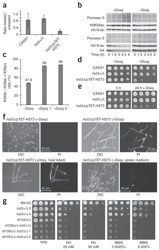
HST3 controls H3K56 deacetylation and is required for cell viability in C. albicans. (a) Repression of HST3 mRNA by doxycycline treatment, as measured by quantitative RT-PCR. Results are the mean of three experiments ± s.e.m. (b) Immunoblots of lysates from hst3Δ/ pTET-HST3 cells treated with doxycycline (+Doxy, 40 μg ml−1) or left untreated (−Doxy). Equal amounts of histone H3 (top) or of total proteins (bottom) were loaded for each time point. (c) Stoichiometry of H3K56ac determined by mass spectrometry of histones purified from hst3Δ/pTET-HST3 cells grown in the absence or presence of doxycycline (duplicates). (d) Growth of hst3Δ/pTET-HST3 cells on plates containing doxycycline (50 μg ml−1) or control plates. CASS1, wild-type strain. (e) Growth of hst3Δ/ pTET-HST3 cells transiently exposed to doxycycline (50 μg ml−1) for 24 h and plated on YPD lacking doxycycline. (f) Lethality of hst3Δ/pTET-HST3 cells caused by doxycycline treatment (20 μg ml−1 for 24 h), as assayed by fluorescence microsopy after propidium iodide staining. Control cells were either heat-killed or triggered to undergo filamention (spider medium). DIC, differential interference contrast. PI, propidium iodide. (g) Susceptibility to genotoxic agents of WT (SN152), single (rtt109Δ/Δ, hst3+/Δ) and double (rtt109Δ/Δ hst3+/Δ, rtt109Δ/Δ hst3Δ/Δ) mutants (tenfold serial dilutions), as determined by colony formation assays.

