Figure 3.

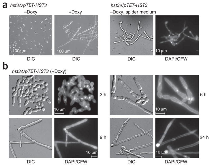
HST3 repression triggers abnormal changes in cell morphology and DNA staining. (a) Left, differential interference contrast (DIC) images of hst3Δ/pTET-HST3 cells grown in YPD medium at 30 °C in the absence or presence of doxycycline (20 μg ml−1 for 24 h). Right, cells grown in spider medium at 37 °C (hyphae-inducing conditions), stained with DAPI and calcofluor white (CFW) to mark DNA and cell walls, respectively, and visualized by epifluorescence microscopy. (b) DIC and epifluorescence images showing the morphology, DNA, septa and cell walls of hst3Δ/pTET-HST3 cells monitored at various times after doxycycline addition.
