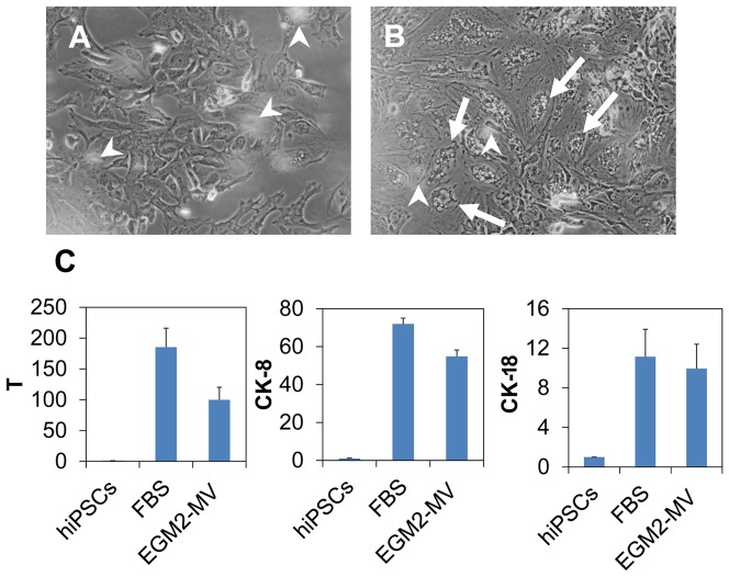
Figure 3. Influence of culture media on the notochordal differentiation.
Two media was investigated: one contained 10% fetal bovine serum (FBS medium) and another contained a cocktail of growth factors (EGM2-MV medium, which is a commercial product of Lonza). (A) and (B) represents the cells differentiated for 5 days in FBS medium and EGM2-MV medium, respectively. Arrow heads indicate the floating NP tissue (out of focus in the image). Frequently the cells (arrows) in B displayed vacuole morphology which is also observable in primary culture of notochordal cells. (C): The transcript levels of three typical notochordal genes of the two cultures. The data are reported in relative mRNA expression which was analyzed by 2−ΔΔCt method using hiPSCs as reference. Three biological samples were pooled and measured so that each result represents the average of three replicates.

