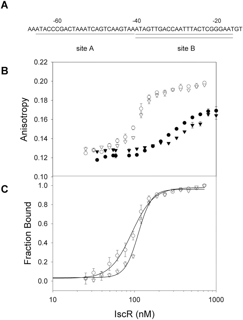
Fig. 4.
Binding isotherms of [2Fe-2S]-IscR and apo-IscR for the two Type 1 sites within the PiscR region. A) Sequences of IscR binding sites A (underlined) and B (double underlined) within PiscR. Numbers indicate the distance relative to the +1 transcription start site. B) DNA binding isotherms of wild-type [2Fe-2S]-IscR (open symbols) and the clusterless mutant protein IscR-C92A/C98A/C104A (closed symbols) measured as a change in anisotropy under anaerobic conditions. Both forms of IscR protein were incubated with 5 nM fluorescently labeled DNA containing either site A (circles) or site B (triangles) in 40 mM Tris-Cl (pH 7.9) and 150 mM KCl. C) Fraction bound corrected for fluorescence quenching of labeled PiscR sites A or B bound by [2Fe-2S]-IscR as a function of protein concentration. Error bars represent the standard errors of triplicate experiments.

