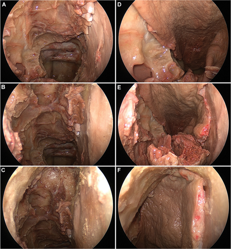
Fig. 4.

Endoscopic endonasal dissection and pictures with 0-degree endoscope. (A–C) Complete anterior and posterior ethmoidectomy, large sphenoidotomy, and frontal sinusotomy (Draf IIb) on the right side for exposure of the entire skull base from the sella to the frontal sinus. (D–F) Rotation and placement of extended inferior turbinate flap with the anterior septal mucosa added.
