Fig. 1.

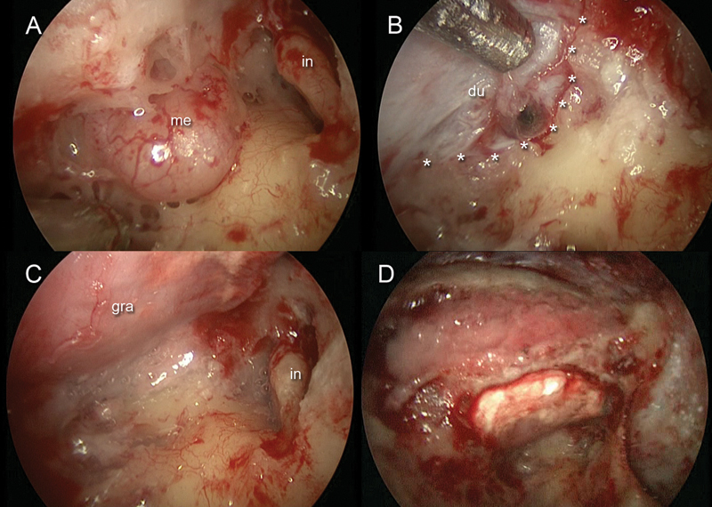
Right ear. (A) Endoscopic view of the meningeal herniation along the mastoid and antral tegmen. (B) After meningeal herniation removal, a 0-degree endoscope was inserted through the mastoid cavity to detect the dural defect and the anterior limit of the tegmental defect (asterisks). (C) A Duragen graft was inserted through the minicraniotomy to repair the defect. (D) Bony dust was placed through the mastoid cavity to cover the tegmental defect and to cover the site of the minicraniotomy. du, dura mater; gra, graft, in, incus; me, meningeal herniation.
