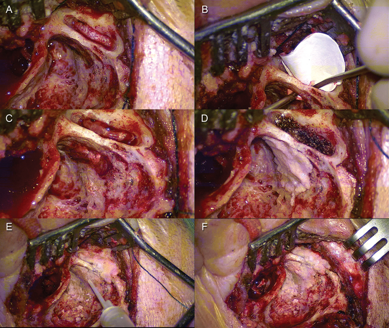
Fig. 2.

Left ear. (A) A minicraniotomy was performed. (B) Duragen graft was positioned through the minicraniotomy. (C) The graft was placed between the dura mater and the superior surface of the middle cranial fossa covering the tegmental defect. (D) Bony dust was placed over the tegmental defect through the mastoid cavity. (E, F) Final cavity after tegmen repair.
