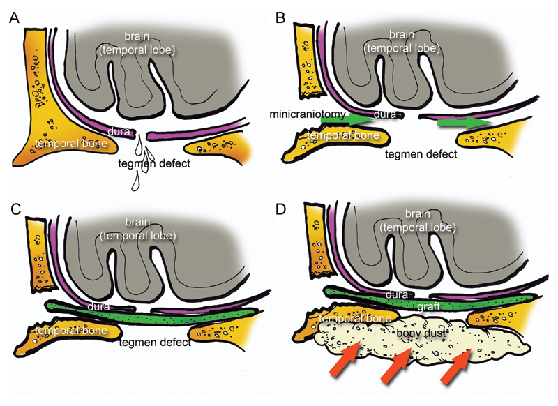
Fig. 3.

Schematic drawing representing the surgical repair with a single layer method. (A) A tegmental defect was detected in association with dura laceration. (B) A minicraniotomy was performed and the dura mater was gently elevated through the minicraniotomy (green arrows) from the superior surface of the middle cranial fossa to create a site for placement of the graft. (C) A graft. (D) Bony dust was placed over the tegmental defect through the mastoid cavity (red arrows).
