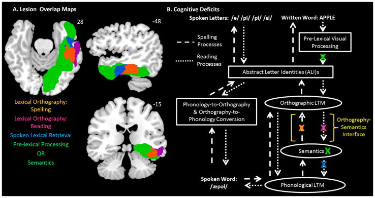
Figure 5.

Cognitive deficits and corresponding lesion maps. (A) Depicts the lesion maps associated with each behavioral pattern depicted in Table 2. The lesion maps are projected onto a standard template brain in MNI coordinate space that was rotated −15 degrees from the AC-PC line. The orange area depicts the overlap of DPT’s, DSN’s, and LHD’s lesions, indicating an area involved in lexical orthographic processing in spelling. The violet area depicts the overlap of DPT’s and DSN’s lesions, excluding LHD’s, indicating an area involved in lexical orthographic processing in reading. The blue area depicts the overlap of DSN’s and LHD’s lesion, excluding DPT’s, indicating an area involved in spoken word retrieval. The green area depicts LHD’s lesion excluding DPT and DSN’s, indicating areas involved in either pre-lexical visual processing or semantic processing/representation. The colored text reports the corresponding cognitive processes associated with each (colored) lesion area. (B) Depicts the theory of written language processing including the proposed deficit locations for DPT, DSN and LHD. Lesions are denoted by a colored X. DPT has impairments in lexical orthography for spelling and reading (orange and violet X’s). DSN has an impairment in lexical orthography for spelling and reading and also an in impairment in spoken lexical retrieval (orange, violet and blue X’s). LHD has an impairment in lexical orthography for spelling, pre-lexical processing, and semantic processing (orange and green X’s). The colored X’s correspond to the specific cognitive deficits described in the text, listed in Table 2 and at the bottom of Figure 5A. The yellow text and bracket indicates the Orthography-Semantics Interface.
