
Figure 1. Clinical Presentation of Proband.
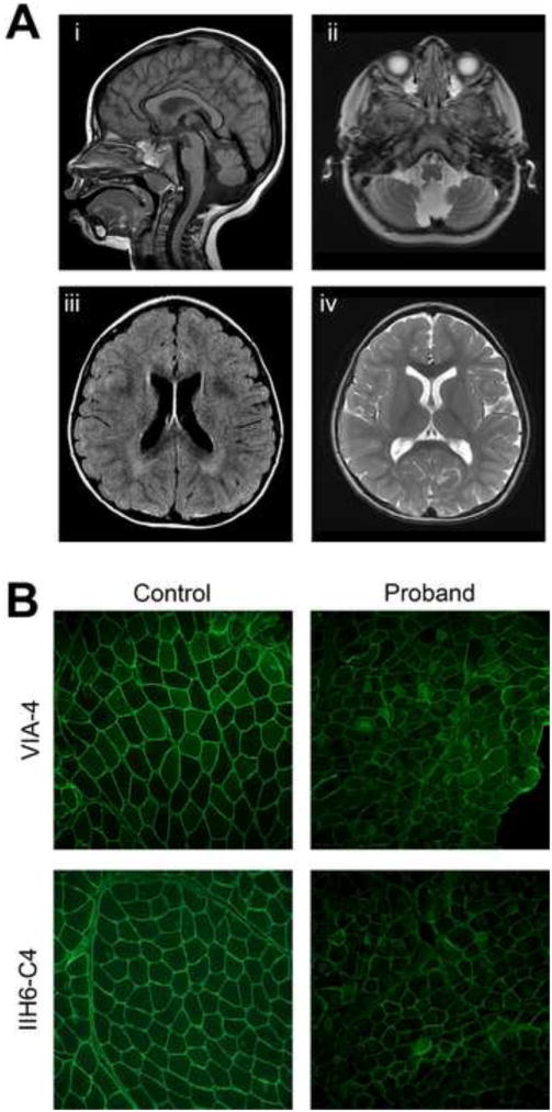
A) Brain MRI of the older child at age 2 years showing hypoplasia of the cerebellar vermis and lower brainstem and cerebellar polymicrogyria on sagittal T1 (i) and axial T2 (ii) views. Axial FLAIR (iii) and axial T2 (iv) images show periventricular white matter T2 hyperintensities suggestive of mild cerebral parenchymal underdevelopment. (B) Immunofluorescence for two epitopes of glycosylated alpha-dystroglycan both show variably diminished staining in the muscle biopsy as compared with control muscle.