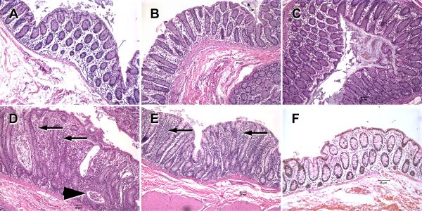
FIGURE 2.
Hematoxylin and Eosin stained tissue sections of colon from acutely SIV-infected rhesus macaques (A- 7DPI), (B and C- 13DPI), and non-SIV-infected macaques with diarrhea (D&E) and a uninfected normal control macaque (F). In the colon of the non-SIV-infected macaques with diarrhea, moderate to severe colitis is evident from diffuse cellular infiltrates (arrows), crypt dilations and crypt abscesses (arrowhead) and a relative paucity of goblet cells (D&E) compared to acutely SIV-infected (A-C) and normal control (F) macaques. Note that the colons of acutely SIV-infected macaques AV91 (A), HI58 (B) and HI63 (C) show minimal to no histological evidence of inflammation. All figures are 10× magnification.

