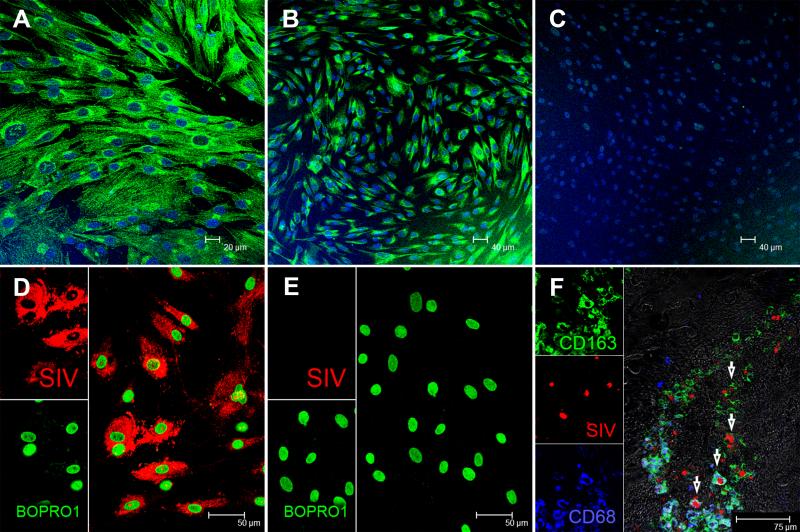
FIGURE 6. Characterization of cultured intestinal macrophages.
In vitro cultured primary intestinal macrophages express classical macrophage markers such as CD68 (A) and CD163 (B) but do not express T cell markers such as CD3 (C). All three panels are double labels with CD68 and CD163 in green and nuclear labeling with Topro3 in blue. Intestinal macrophages were infected with SIVmac251 and in situ hybridization confirmed the presence of viral RNA (D). Uninfected cells are negative for SIV (E). Both panels involve double labels with viral RNA (red) and Bopro1 (green) for nuclear staining. SIV-infected macrophages can be detected in the intestine as early as 10 days post SIV infection (F). Arrows point to SIV-infected (red) macrophages that express CD68 (blue), CD163 (green) or both.

