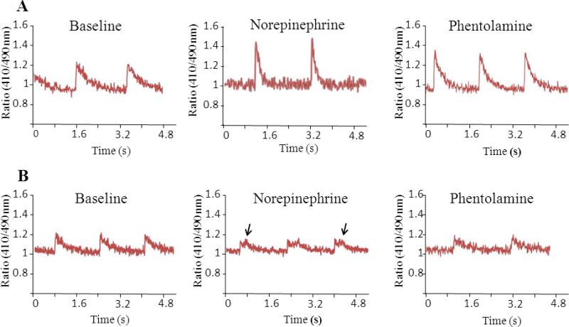
Figure 3.
Calcium transients in cardiomyocytes isolated from wild type (A) and CASQ2Δ/Δ (B) mice. Representative traces are shown on baseline, after exposure to norepinephrine and then after adding phentolamine. Note the repetitive abnormal calcium release waves in CASQ2Δ/Δ cells exposed to norepinephrine (arrows) which were seen in 4/12 cells studied and disappeared after adding phentolamine (mean±SD, n=3-5 mice/group).

