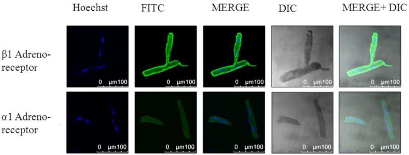
Figure 6.
Confocal microscopy in an isolated CASQ2Δ/Δ cardiomyocyte. Cells were stained for either 1adreno-receptor (upper panels) or for 1 adreno-receptor (lower panel).Abbreviations: Hoechst, nucleus staining; FITC, green fluorescence staining; MERG, Hoechst+ FITC; DIC- light microscopy; MERG+ DIC, Hoechst+ FITC+ light microscopy.

