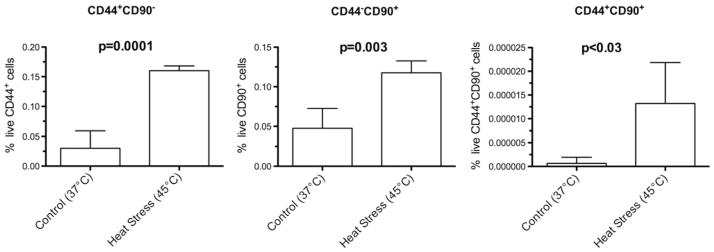
Fig. 2.

Effect of sublethal heat stress on proportion of live CD44+ , CD90+ , and CD44+ CD90+ N1S1 HCC cells. Graph shows that sublethal heat stress (45 °C, 10 min) resulted in significant increase in proportion of live CD44+ CD90− (5.3-fold), CD44−CD90+ (2.4-fold), and CD44+ CD90+ (22.0-fold) N1S1 HCC cell subpopulations relative to control group (37 °C, 10 min) after 48-h recovery. Treatment groups compared by unpaired t test. Data are presented as mean ± SEM from 4 independent cell cultures
