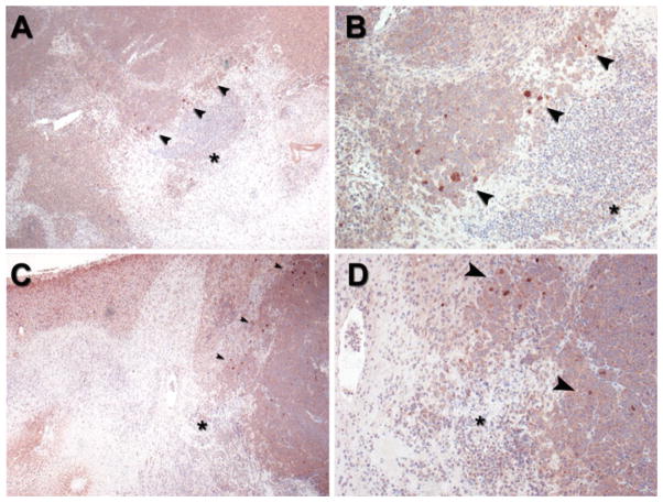
Fig. 5.
Representative CD44 immunostaining at tumor ablation margin in orthotopic N1S1 HCC tumor model from 2 different rats (A, B and C, D, respectively). A, C Low-power (40×) and B, D high-power (100×) photomicrographs of ablation zone (ablated tissue denoted by black asterisk) from both rats demonstrate clusters of rare cells staining positive for CD44 (brown) at tumor ablation margin (black arrowheads) 24 h after partial laser ablation

