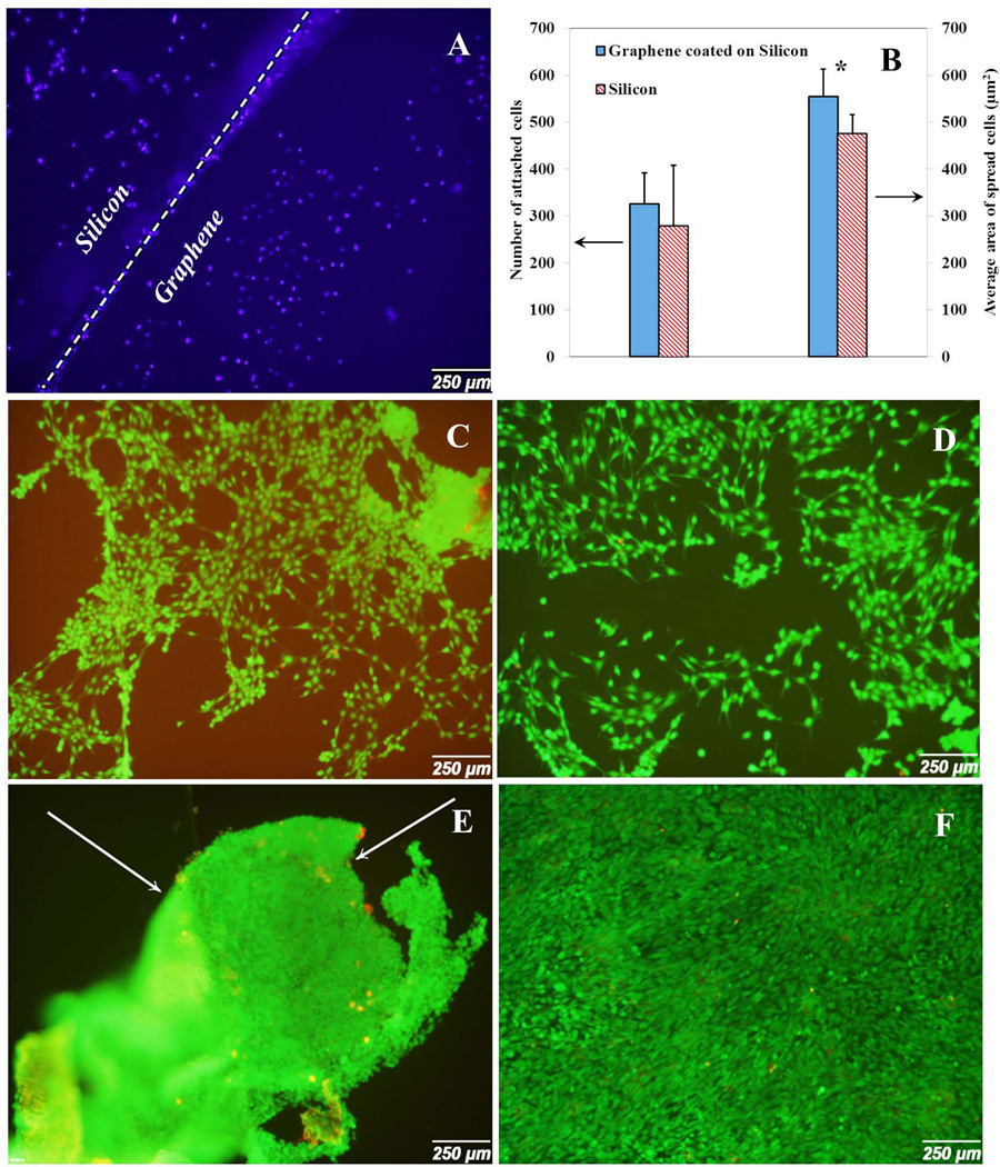
Fig. 7.
Cell attachment and proliferation on silicon wafer substrate with and without graphene coated layer at day 2 and day 5. (A) DAPI stained the cell nucleus at the border of graphene and silicon at day 2. (B) Number of attached cells and average area of spread cells at day 2 for silicon wafer with and without graphene layer. (C) Cell spreading on silicon substrate at day 2. (D) Cell spreading on graphene coated silicon wafer substrate at day 2. (E) Cell spreading on silicon wafer substrate at day 5. (F) Cell spreading on graphene coated silicon wafer substrate at day 5. Arrows show the detachment of cell layer from the substrate.

