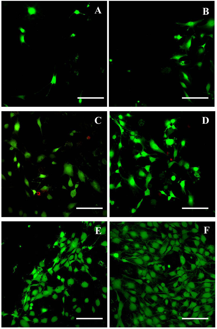
Fig. 9.
High magnification images to show cell morphology on different substrates at day 2. (A) Glass substrate without graphene coated layer. (B) Graphene coated glass substrate (C) Silicon wafer substrate without graphene layer. (D) Graphene coated silicon wafer substrate. (E) Stainless steel without graphene coated layer. (F) Graphene coated Stainless steel substrate. Scale bar=200 µm.

