Figure 2.

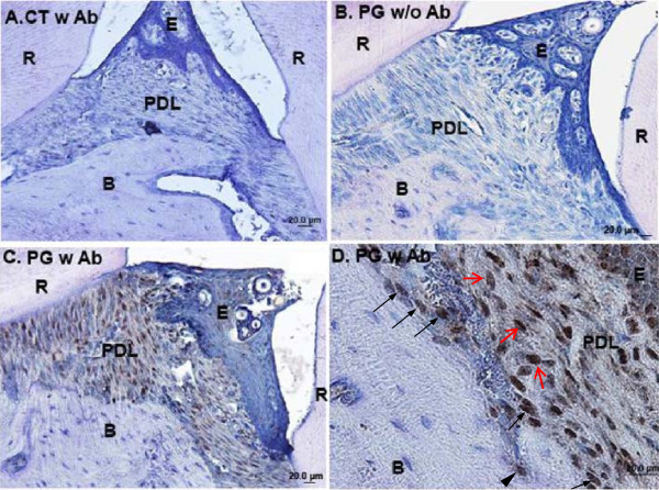
P. gingivalis invade periodontal soft and hard tissues after repetitive inoculations as shown by immunohistochemistry. Immunohistochemistry was performed three days after three times of P. gingivalis inoculations. A. Sham-infected control. Anti-P. gingivalis primary antibody was applied in the assay. Blue is hematoxylin counterstaining. Notice there is no brown positive staining for P. gingivalis in the periodontium. B. P. gingivalis infected animals, with anti-P. gingivalis primary antibody excluded from the assay, which shows no positive staining in the periodontium. This demonstrated there was no unspecific staining from secondary antibody and/or substrate. C. P. gingivalis infected animals, with anti-P. gingivalis primary antibody included in the assay. Extensive staining for P. gingivalis was noticed in gingival epithelial cells, PDL fibroblasts, and alveolar osteoblasts. D. Magnified view in C. Positive P. gingivalis staining was detected in fibroblasts (denoted by red arrows) in periodontal ligament space, osteoblasts lining the alveolar bone surface (denoted by black arrows), and in an osteocyte embedded in the alveolar bone matrix (denoted by black arrow head). Abbreviations: CT, control, sham-infected; PG, P. gingivalis infected; Ab, antibody; R, root; E, gingival epithelial cells; PDL, periodontal ligament; B, alveolar bone; scale bar = 20 μm.
