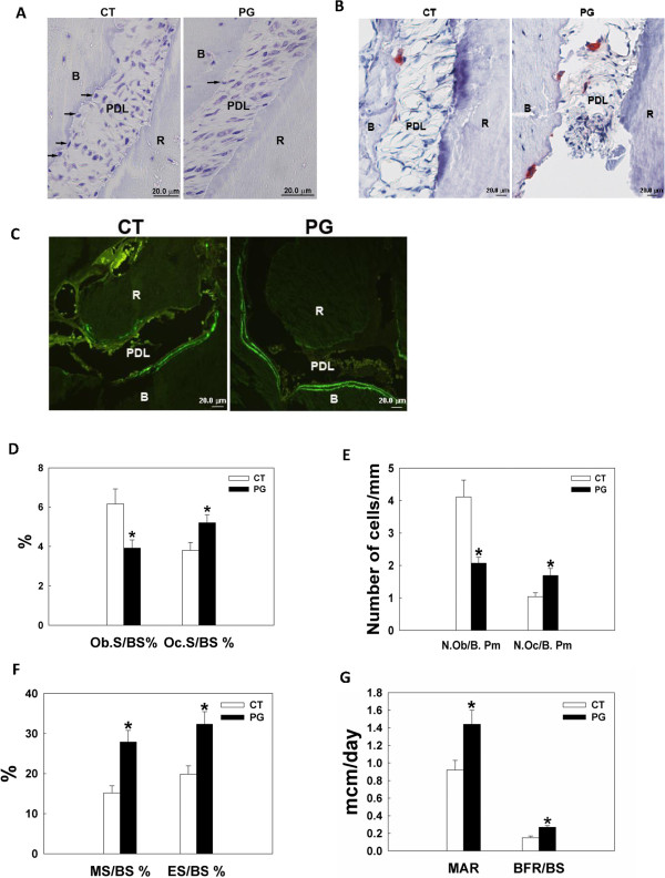
Figure 4.

P. gingivalis infection results in increased osteoclastic bone resorption and increased osteoblastic bone formation as shown by bone histomorphometric analysis. Bone histomorphometry was done four weeks after a total of eight bacterial inoculations. A. Toluidine blue staining for osteoblasts. Arrows denote osteoblasts in contact with the alveolar bone surface. B. TRAP staining for osteoclasts, which are red-stained multinucleated cells. C. Calcein doubling labeling to show newly formed bone. The area between the double bright green lines is where the new bone is formed. D-G, quantified bone histomorphometry data. A significant decrease of osteoblast number (D and E), increased osteoclast number (D and E), and increased osteoclastic bone resorption (F) was observed in the infected animals. Surprisingly, osteoblastic bone formation was greatly elevated in the remaining osteoblasts (F and G). Abbreviations: CT, control, sham-infected; PG, P. gingivalis infected; B, alveolar bone; R, root; PDL, periodontal ligament; Ob.S/BS%, percent bone surface lined with osteoblasts; Oc.S/BS%, percent bone surface covered with osteoclasts; N.Ob/B. Pm, number of osteoblasts per bone perimeter; N. Oc/B. Pm, number of osteoclasts per bone perimeter; MS/BS%, percent of mineralizing surface of total bone surface measured; ES/BS%, percent bone surface eroded by osteoclasts; MAR, mineral apposition rate; BFR/BS, bone formation rate; *, denotes P < 0.05 compared with the controls. Scale bar = 20 μm.
