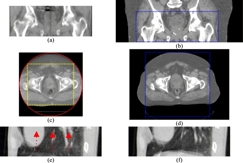
FIG. 1.
Examples of images partially matching situations. (a)–(d) are the scans of a prostate cancer patient. (a) and (c) are tomotherapy MVCT scans. (b) and (d) are the corresponding conventional kVCT scans. (a) and (b) demonstrate the different superior-inferior coverage between the MVCT and kVCT. The dashed lines in (b) and (d) mark the boundaries of the corresponding MVCT volume. (c) and (d) demonstrate the differences in FOV. The circle in (c) is the tomotherapy MVCT FOV circle. The rectangle marks the maximal cropped region if the MVCT image is cropped to be completely within the FOV. (e) and (f) are two 64-slice axial CT lung scans at the same couch position but at different phases of the breathing cycle. The arrows in (e) demonstrate the directions of tissue motion to match the scan in (f).

