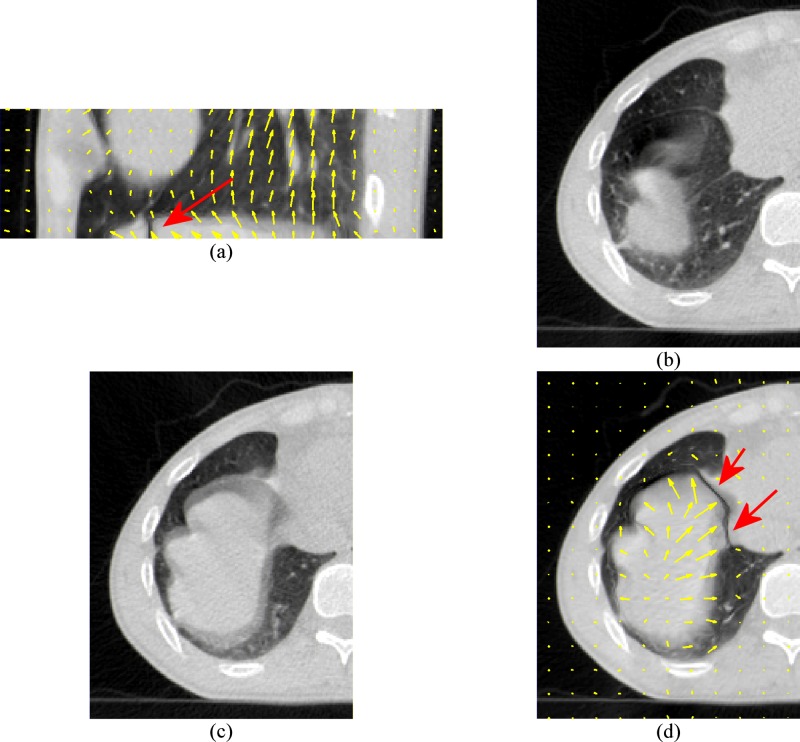
FIG. 2.
Demonstration of registration problems on the 64-slice lung CT scans. The CT scan in Fig. 1(e) is used as the moving image and the CT scan in Fig. 1(f) is used as the fixed image. (a) and (d) are the deformed moving image. (b) is the moving image in transverse view. (c) is the fixed image in transverse view. The arrows indicate the most significant registration errors. The motion vectors in (a) and (d) also show the incorrect transverse motions as the diaphragm should move mainly in the superior-inferior direction instead of in the transverse direction.

