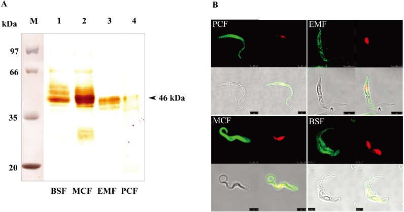
Fig. 3.
Detection of the native TcP46 from all life cycle stages of the parasite. (A) Lane M: Molecular size marker. Western blot analysis of the native TcP46 was carried out using the cell lysate from BSF, MCF, EMF and PCF stages of T. congolense and anti-rTcP46 mouse serum. (B) Cellular localizations of the TcP46 in all four life cycle stages of T. congolense (PCF, EMF, MCF and BSF) were examined by immunofluorescence staining and confocal laser scanning microscopy. Green indicates immunofluorescence staining of TcP46, and red indicates nucleus and kinetoplast.

