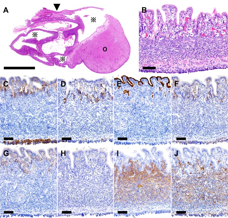
Fig. 1.
Right ovary. (A) A large cyst (※) in the ovarian hilus (arrowhead) is observed. (B) The cyst wall is lined with monolayer cuboidal epithelial cells with cilia. Beneath the epithelial layer, luteinized stromal cells and spindle-shaped stromal cells of the ovary are observed. (C) All the epithelial cells are positive for pan-CK. (D) Epithelial cells invaginating into the stroma are positive for CK18. (E) The apical surface of the epithelial cells is positive for CD10. (F) Nuclei of the epithelial cells are positive for estrogen receptor. (G) Nuclei and the cytoplasms of the epithelial cells are positive for calretinin. (H) No SMA-positive muscular layer is observed beneath the epithelial layer. (I) Luteinized stromal cells and spindle-shaped stromal cells beneath the epithelial layer are positive for vimentin. (J) Luteinized stromal cells and spindle-shaped stromal cells beneath the epithelial layer are positive for melan A. Arrowhead, ovarian hilus; ※, cyst; O, ovary. (A and B) HE; (C–J) immunohistochemistry. (A) Bar=5 mm; (B–J) bar=50 µm.

